
Design of pulse laser coupling scheme for observing channels through hysteroscopy
-
摘要: 为了提高脉冲激光通过宫腔镜的能量,采用相关软件设计了3种透镜耦合方案来提高耦合效率,进行了理论分析和实验验证。结果表明,耦合效率约提高了14%,脉冲激光通过该耦合模块后能取得较好的光声成像效果。该方法可作为辅助工具应用于临床中子宫内膜等疾病的检测,有望提高光声成像技术在子宫内膜的应用。Abstract: In order to improve the energy of the pulsed laser passing through the hysteroscope, three lens coupling scheme was adopted was adopted to improve the coupling efficiency. The theoretical analysis and experimental validation were carried out. The results show that the coupling efficiency is increased by about 14%. The experimental data, which indicates that the pulsed laser is more powerful than the pulsed laser, is more efficient than the pulsed laser. A better photoacoustic imaging effect is obtained after passing through our proposed coupling module. The study is expected to improve the effect of photoacoustic imaging technology in endometrial application, and will be able to be applied as an auxiliary tool for the detection of endometrial and other diseases in the clinic.
-
Keywords:
- medical optics and biotechnology /
- pulsed laser /
- lens coupling /
- hysteroscope
-
-
![]()
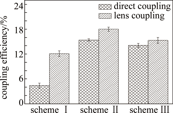
图 8 直接耦合与透镜耦合效率对比图
Figure 8. Comparison of efficiency between direct coupling and lens coupling
![]()
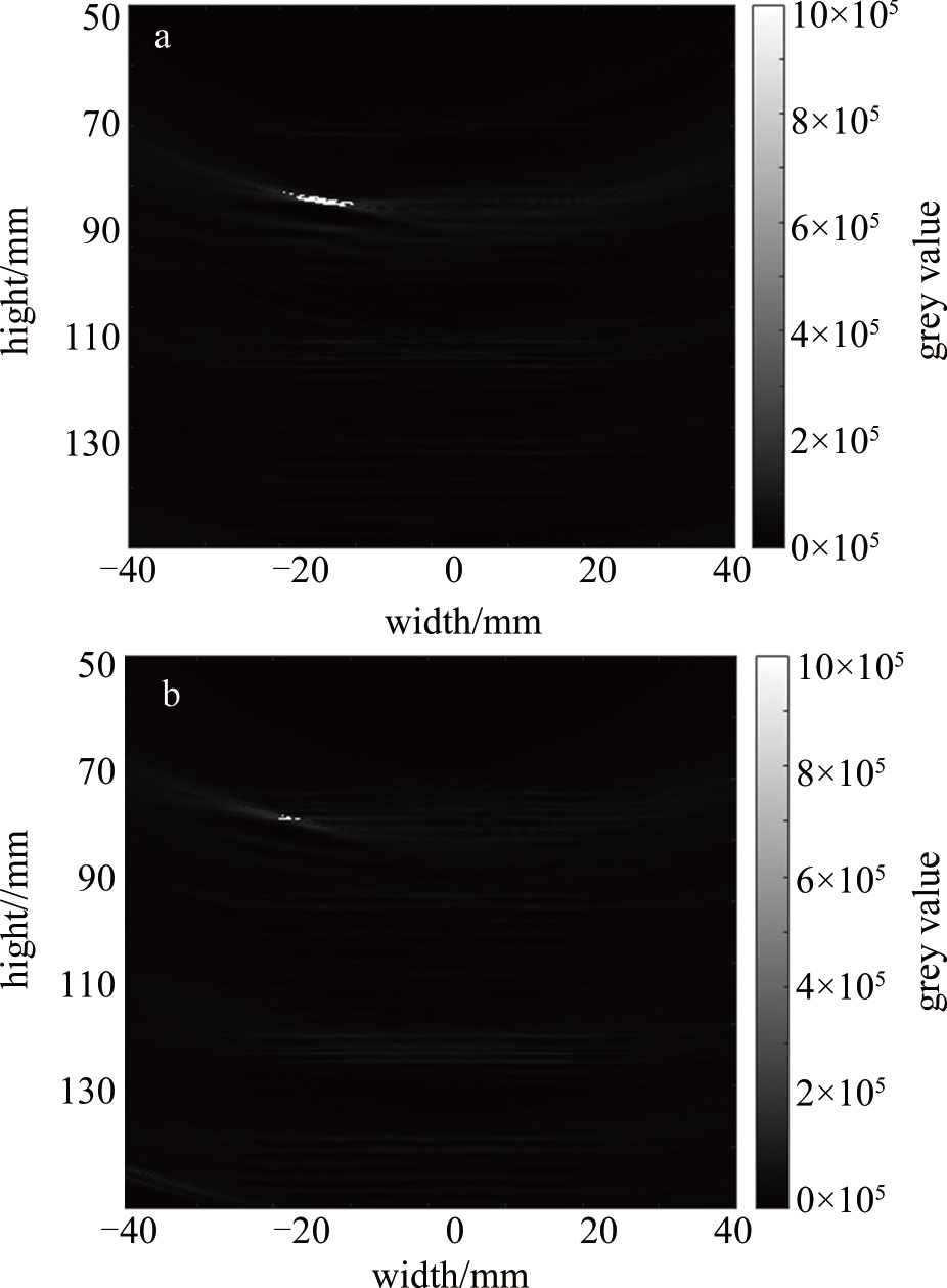
图 10 a—透镜耦合光声信号图b—直接耦合光声信号图
Figure 10. a—lens coupled photoacoustic signal diagram b—direct coupled photoacoustic signal diagram
表 1 耦合系统参数
Table 1 Coupling system parameter
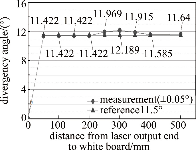
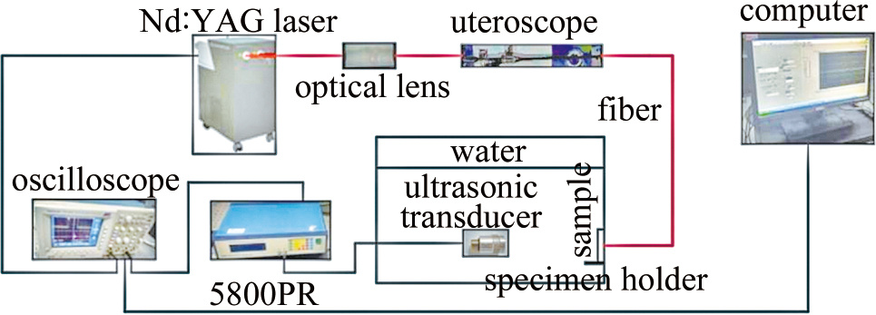
coupling system parameter indicators laser model PhotoSonus model YAG laser pulse laser wavelength 660 nm laser divergence angle 11.5° hysteroscopy model direct tube hysteroscopy produced by TSCS hysteroscopy diameter 6.5 mm lens 1 f1=20 mm, coated lens 2 f2=-15 mm, coated  下载: 导出CSV
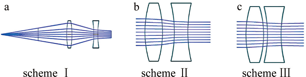
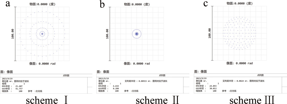
表 2 3种耦合方案具体参数
Table 2 Specific parameters of three coupling schemes
pulsed laser avelength/nm lens focal length/mm optical center distance/mm schemeⅠ 660 f1=20, f2=-15 19.644 scheme Ⅱ 6.09 scheme Ⅲ 4.31
下载: 导出CSV
-
[1] 魏建磊, 张涛, 张鹏霞. 治疗性肿瘤疫苗: 过去、现在和未来[J]. 中国生物化学与分子生物学报, 2023, 39(10): 1-14. WEI J L, ZHANG T, ZHANG P X. Therapeutic tumor vaccines: Past, present and future[J]. Chinese Journal of Biochemistry and Molecular Biology, 2023, 39(10): 1-14(in Chinese).
[2] MARKAROV A E, APRESYAN S V, DIMITROVA V I, et al. The choice of the X-ray endovascular method for the treatment of uterine fibroids and the possibility of prolonging the remission of the disease[J]. Meditsinskiy Sovet, 2021(13): 51-56. DOI: 10.21518/2079-701X-2021-13-51-66
[3] LIU J Y, CHANG C, ZHANG H X. Grayscale ultrasound feature typing of metastatic ovarian tumors, particularly signet-ring cell carcinoma[J]. Quantitative Imaging in Medicine and Surgery, 2023, 13(1): 49-57. DOI: 10.21037/qims-21-1149
[4] TURASHVILI G, HANLEY K. Practical updates and diagnostic cha-llenges in endometrial carcinoma[J]. Archives of Pathology & Laboratory Medicine, 2023, 148(1): 78-98.
[5] 杨永秀. 妇科肿瘤发病机制及治疗的研究与应用[EB/OL]. (2020-11-16)[2023-10-16]. https://ir.lzu.edu.cn/handle/262010/562735. YANG Y X. Research and application of pathogenesis and treatment of gynecologic tumors[EB/OL]. (2020-11-16)[2023-10-16]. https://ir.lzu.edu.cn/handle/262010/562735 (in Chinese).
[6] 杨苗苗. 宫腔镜检查与早期子宫内膜癌腹腔冲洗液细胞学及预后的相关性分析[D]. 郑州: 郑州大学, 2020: 14-17. YANG M M. Correlation analysis of hysteroscopy with cytology and prognosis of abdominal lavage fluid in early endometrial cancer[D]. Zhengzhou: Zhengzhou University, 2020: 14-17 (in Chinese).
[7] OYAMA S, TANAKA K, MORIYAMA M, et al. Laparoscopic resection of an intra-abdominal esophageal duplication cyst in the ileum: A case report[J]. Surgical Case Reports, 2022, 8(1): 219. DOI: 10.1186/s40792-022-01576-6
[8] MEENAKSHI G, AASMA N, PRATIBHA S, et al. Role of cervical cancer biomarkers p16 and Ki67 in abnormal cervical cytological smear[J]. The Journal of Obstetrics and Gynecology of India, 2021, 71(1): 72-77. DOI: 10.1007/s13224-020-01380-y
[9] NGUYEN D B, GERBER V E M, SUEN M W H, et al. Outpatient hysteroscopy is effective for uterine cavity evaluation following failed office-based endometrial biopsy[J]. The Journal of Obstetrics and Gynaecology Research, 2022, 48(9): 1.
[10] 冯肖媛, 陶旭炜, 曾凌空, 等. 肺部超声在新生儿新型冠状病毒肺炎诊断中的应用[J]. 中华儿科杂志, 2020, 58(5): 347-350(in Chinese). FENG X Y, TAO X W, ZENG L K, et al. Application of pulmonary ultrasound in the diagnosis of COVID-19 pneumonia in neonates[J]. Chinese Journal of Pediatrics, 2020, 58(5): 347-350.
[11] 廖涛, 龚金玲, 付赤学. 经阴道超声造影诊断早期子宫内膜癌的临床价值[J]. 武警医学, 2022, 33(9): 751-754. LIAO T, GONG J L, FU Ch X. Clinical value of transvaginal ultrasonography in diagnosing early endometrial cancer[J]. Armed Police Medicine, 2022, 33(9): 751-754 (in Chinese).
[12] 岳巍. 多模型扩散加权成像对子宫内膜癌的诊断及其预后因素的相关性分析[D]. 郑州: 郑州大学, 2022: 72-78. YUE W. Correlation analysis of multi-model diffusion-weighted imaging for the diagnosis of endometrial cancer and its prognostic factors[D]. Zhengzhou: Zhengzhou University, 2022: 72-78(in Chinese).
[13] 邵迎华, 杨秀梅, 刘洋, 等. 宫腔镜与超声对子宫内膜病变诊断的对比研究[J]. 中国医学设备, 2022, 19(7): 103-106. SHAO Y H, YANG X M, LIU Y, et al. Comparative study of endometriosis diagnosis by hysteroscopy and ultrasound[J]. China Medical Equipment, 2022, 19(7): 103-106(in Chinese).
[14] 穆根, 张振辉, 石玉娇. 生物医学影像中的光声成像技术[J]. 中国激光, 2022, 49(20): 2007208. MU G, ZHANG Zh H, SHI Y J. Photoacoustic imaging in biomedical imaging[J]. Chinese Journal of Lasers, 2022, 49(20): 2007208 (in Chinese).
[15] YANG L, LI Y P, FANG F, et al. Highly sensitive and miniature microfiber-based ultrasound sensor for photoacoustic tomography[J]. Opto-Electronic Advances, 2022, 5(6): 200076. DOI: 10.29026/oea.2022.200076
[16] 李长辉. 用光奏响生命之歌: 光声成像技术漫谈[J]. 激光与光电子学进展, 2022, 59(6): 0617005. LI Ch H. Creating the sound of life by light: A discussion about photoacoustic imaging[J]. Laser & Optoelectronics Progress, 2022, 59(6): 0617005(in Chinese).
[17] CHU B B, CHEN Zh M, SHI H L, et al. Fluorescence, ultrasonic and photoacoustic imaging for analysis and diagnosis of diseases[J]. Chemical Communications, 2023, 59(17): 2399-2412. DOI: 10.1039/D2CC06654H
[18] 徐大宝, 冯力民. 宫腔镜手术技巧及并发症防治[M]. 北京: 人民卫生出版社, 2019: 15-18. XU D B, FENG L M. Hysteroscopic surgical techniques and prevention of complications[M]. Beijing: People's Health Publishing House, 2019: 15-18(in Chinese).
[19] SMIT J G, OVERDIJKINK S, MOL B W, et al. The impact of diagnostic criteria on the reproducibility of the hysteroscopic diagnosis of the septate uterus: A randomized controlled trial[J]. Human Reproduction, 2015, 30(6): 1320-1330.
[20] 段华. 中国妇科内镜诊疗技术从弱到强并跑国际之路[J]. 中国计划生育和妇产科, 2019, 11(10): 3-6. DUAN H. China' s gynecologic endoscopy diagnosis and treatment technology from weak to strong and run the international road[J]. China Family Planning and Obstetrics and Gynecology, 2019, 11(10): 3-6(in Chinese).
[21] 郑荣寿, 孙可欣, 张思维, 等. 2015年中国恶性肿瘤流行情况分析[J]. 中华肿瘤杂志, 2019, 41(1): 19-28. ZHENG R S, SUN K X, ZHANG S W, et al. Analysis of the prevalence of malignant tumors in China in 2015[J]. Chinese Journal of Oncology, 2019, 41(1): 19-28 (in Chinese)
[22] 马立芳, 贾岚, 李强. 子宫内膜癌的实验室及影像学指标的检测意义——评《子宫内膜癌100问》[J]. 中国实验方剂学杂志, 2022, 28(24): 34. MA L F, JIA L, LI Q. Significance of laboratory and imaging indicators for the detection of endometrial cancer—a review of 100 questions on endometrial cancer[J]. Chinese Journal of Experimental Formulary, 2022, 28(24): 34(in Chinese).
[23] 田源. 经阴道超声联合宫腔镜检查对子宫内膜癌的诊断价值[J]. 中国当代医药, 2022, 29(15): 101-103. TIAN Y. Diagnostic value of transvaginal ultrasound combined with hysteroscopy in endometrial cancer[J]. China Contemporary Medicine, 2022, 29(15): 101-103 (in Chinese).
[24] 林永平. 面向早期子宫肿瘤诊断的光声与超声双模成像原理与技术[D]. 福州: 福建师范大学, 2019: 32- 48. LIN Y P. Principles and techniques of photoacoustic and ultrasound dual-mode imaging for early uterine tumor diagnosis[D]. Fuzhou: Fujian Normal University, 2019: 32- 48(in Chinese).
[25] 唐华, 沈咏, 龙丽媛. 国家自然科学基金视角下我国激光科学技术发展的分析和展望[J]. 中国激光, 2023, 50(2): 0200001. TANG H, SHEN Y, LONG L Y. Analysis and prospect of the development of laser science and technology in china from the perspective of national natural science foundation of China[J]. Chinese Journal of Lasers, 2023, 50(2): 0200001 (in Chinese).
-
期刊类型引用(3)
1. 张艳红,卢腾飞,刘永欣,陈子阳,孙顺红. 非均匀偏振光束在海洋湍流中的光强特性. 激光技术. 2020(03): 310-314 .  本站查看
本站查看
2. 周正兰,袁扬胜,束杰,徐翔,屈军. 部分相干月牙形光束在非Kolmogorov谱中的漂移. 激光技术. 2019(04): 143-148 . 本站查看
3. 包训旺,袁扬胜,崔执凤,屈军. 受遮挡贝塞尔-高斯光束在湍流大气传输的M~2因子. 激光技术. 2018(03): 427-432 . 本站查看
其他类型引用(2)

计量
- 文章访问数: 26
- HTML全文浏览量: 5
- PDF下载量: 28
- 被引次数: 5