Progresses of surface enhanced fluorescence
-
摘要: 在外光场激励下,金属纳米结构衬底表面所形成的集体电子振荡模式可有效调制其局域电磁场分布,对居于衬底附近的荧光分子的荧光辐射产生调控。其影响因素主要取决于衬底金属表面所形成的电磁振荡模式和电磁场分布性质。归纳了光谱学中表面增强荧光效应研究的关键问题,指出了周期性有序衬底金属增强荧光及其金属纳米颗粒增强荧光研究的主要研究进展。基于局域电磁场增强机理模型,讨论了不同形貌衬底金属对荧光分子的荧光调控机理和影响因素。对表面增强荧光效应的研究前景进行了展望。Abstract: Under the excitation of the external light field, the collective electron oscillation mode formed on the surface of the metal nanostructure can effectively modulate the local electromagnetic field distribution, and control the fluorescence radiation of fluorescent molecules near the substrate. The affecting factors mainly depend on electromagnetic oscillation mode and electromagnetic field distribution formed on the surface of the substrate. The key problems in the study on surface enhanced fluorescence effect in spectroscopy are summarized. The main progress of the research of periodically ordered substrate metal enhanced fluorescence and metal nanoparticle enhanced fluorescence is performed. Based on local electromagnetic field enhancement mechanism model, the mechanism and affecting factors of fluorescence regulation of fluorescent molecules on different substrates are discussed. The research prospect of surface enhanced fluorescence effect is prospected.
-
-
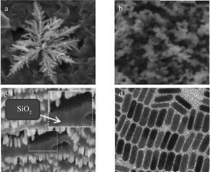
图 5 SiF与AgNP构成的等离激元复合纳米结构[55]
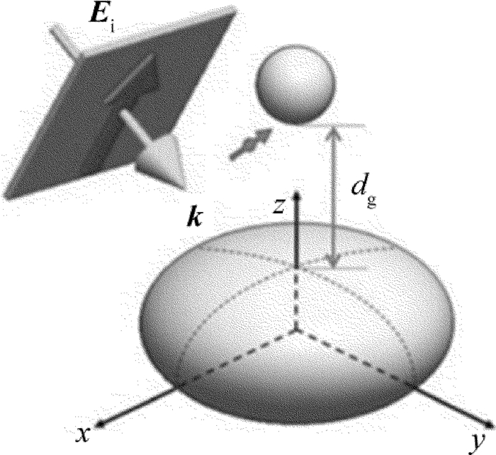
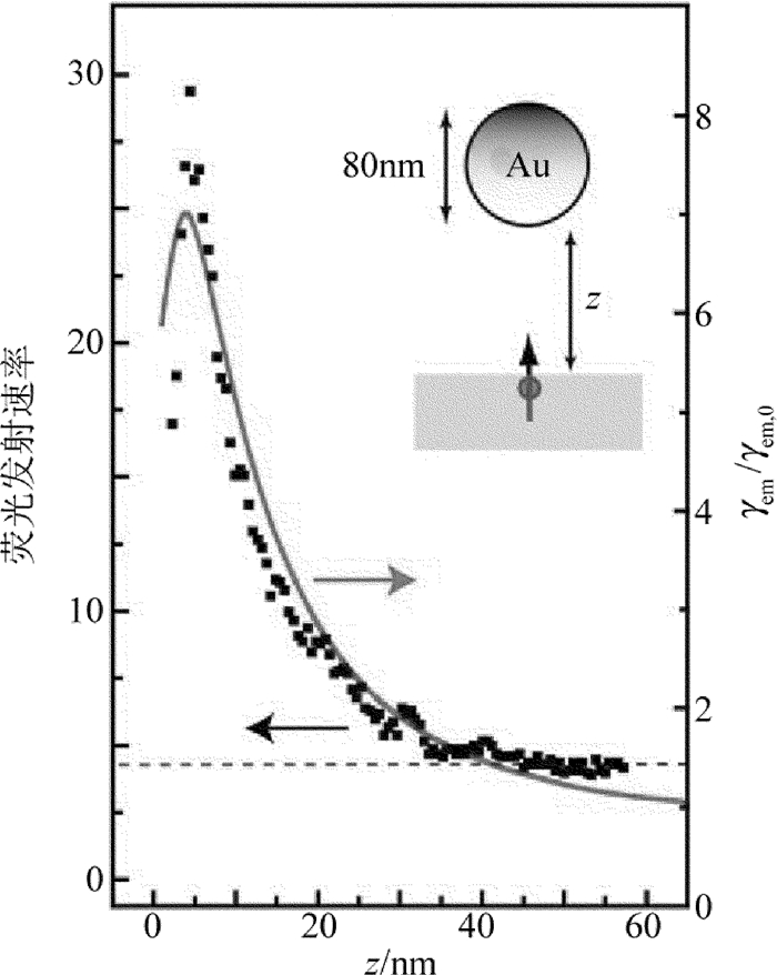
图 8 荧光发射速率随衬底金属到发光中心距离的变化关系[72]
-
[1] DONG J, ZHANG Zh L, ZHENG H R, et al. Recent progress on plasmon-enhanced fluorescence[J]. Nanophotonics, 2015, 4(1):472-490. http://www.wanfangdata.com.cn/details/detail.do?_type=perio&id=nanoph-2015-0028
[2] CHE Y L, CAO X L, LI Q Sh. Preparation and photoluminescence of nano-porous oxidized silicon[J]. Laser Technology, 2008, 32(6):605-607(in Chinese). http://www.wanfangdata.com.cn/details/detail.do?_type=perio&id=jgjs200806004
[3] XU L M, ZHANG Zh L, ZHENG H R, et al. Physical mechanisms of fluorescence enhancement on metal surface[J]. Chinese Journal of Luminescence, 2009, 30(3):8-12(in Chinese).
[4] DREXHAGE K H. Influence of a dielectric interface on fluorescence decay time[J]. Journal of Luminescence, 1970, 1/2:693-701. DOI: 10.1016/0022-2313(70)90082-7
[5] GERSTEN J, NITZAN A. Spectroscopic properties of molecules interacting with small dielectric particles[J]. Journal of Chemical Phy-sics, 1981, 75(3):1139-1152. DOI: 10.1063/1.442161
[6] WEITZ D A, GAROFF S, GERSTEN J I, et al. The enhancement of Raman scattering, resonance Raman scattering and fluorescence from molecules adsorbed on a rough silver surface[J]. Journal of Chemical Physics, 1983, 78(9):5324-5338. DOI: 10.1063/1.445486
[7] GEDDES C D, LAKOWICZ J R. Metal-enhanced fluorescence[J]. Journal of Fluorescence, 2002, 12(2):121-129. DOI: 10.1023/A:1016875709579
[8] LAKOWICZ J R, SHEN Y B, D'AURIA S, et al. Radiative decay engineering:2. Effects of silver island films on fluorescence intensity, lifetimes, and resonance energy transfer[J]. Analytical Biochemistry, 2002, 301(2):261-277. DOI: 10.1006/abio.2001.5503
[9] LÜ G W, LI W Q, ZHANG T Y, et al. Plasmonic-enhanced molecular fluorescence within isolated bowtie nano-apertures[J]. American Chemical Society Nano, 2012, 6(2):1438-1448. http://d.old.wanfangdata.com.cn/NSTLQK/NSTL_QKJJ0225754288/
[10] LÜ G W, SHEN H M, CHENG Y Q, et al. Advances in localized surface plasmon enhanced fluorescence[J]. Science Bulletin, 2015, 60(33):3169-3179(in Chinese). http://www.wanfangdata.com.cn/details/detail.do?_type=perio&id=4a31f30995b415e95ab411a621981256
[11] DONG J, ZHENG H R, LI X Q, et al. Surface-enhanced fluorescence from silver fractallike nanostructures decorated with silver nanoparticles[J]. Applied Optics, 2011, 50(31):123-126. DOI: 10.1364/AO.50.00G123
[12] ZHENG H R, DONG J, ZHANG Zh L, et al. Controlling on the luminescence property of RE ionic and molecular optical centers with nanostructure system[J]. Science Bulletin, 2015, 60(10):899-911(in Chinese). DOI: 10.1007/s11434-015-0785-0
[13] CUI X Q, TAWA K, HORI H, et al. Tailored plasmonic gratings for enhanced fluorescence detection and microscopic imaging[J]. Advanced Functional Materials, 2010, 20(4):546-553. DOI: 10.1002/adfm.v20:4
[14] HAO Q, DU D Y, WANG C X, et al. Plasmon-induced broadband fluorescence enhancement on Al-Ag bimetallic substrates[J]. Scientific Reports, 2014, 4:6014. http://europepmc.org/articles/PMC4127495
[15] TIAN R, YAN D P, LI Ch Y, et al. Surface-confined fluorescence enhancement of au nanoclusters anchoring to a two-dimensional ultrathin nanosheet toward bioimaging[J]. Nanoscale, 2016, 8(18):9815-9821. DOI: 10.1039/C6NR01624C
[16] YANG J, SONG J T, HU W, et al. Fabrication of fluorescence enhancement of quantum dots on a gold colloid formed film for oligonucleotide DNA detection[J]. Analytical Methods, 2017, 9(3):434-442. DOI: 10.1039/C6AY02514E
[17] BROLO A G, KWOK S C, MOFFITT M G, et al. Enhanced fluorescence from arrays of nanoholes in a gold film[J]. Journal of the American Chemical Society, 2005, 127(42):14936-14941. DOI: 10.1021/ja0548687
[18] ZHANG Zh L, ZHENG H R, LIU M C, et al. Surface enhanced fluorescence of Rh6G with gold nanohole arrays[J]. Journal of Nanoscience and Nanotechnology, 2011, 11(11):9803-9807. DOI: 10.1166/jnn.2011.5305
[19] ZHANG Q W, WU L, WONG T I, et al. Surface plasmon-enhanced fluorescence on aunanohole array for prostate-specific antigen detection[J]. International Journal of Nanomedicine, 2017, 12:2307-2314. DOI: 10.2147/IJN
[20] HUNG Y J, SMOLYANINOV I I, DAVIS C C, et al. Fluorescence enhancement by surface gratings[J]. Optics Express, 2006, 14(22):10825-10830. DOI: 10.1364/OE.14.010825
[21] CHOU R Y, LI G T, CHENG Y Q, et al. Surface enhanced fluorescence by metallic nano-apertures associated with stair-gratings[J]. Optics Express, 2016, 24(17):19567-19573. DOI: 10.1364/OE.24.019567
[22] JIANG Y, WANG H Y, SUN H B, et al. Surface plasmon enhanced fluorescence of dye molecules on metal grating films[J]. Journal of Physical Chemistry, 2011, C115(25):12636-12642. http://cpfd.cnki.com.cn/Article/CPFDTOTAL-GXJM201106001131.htm
[23] YIN Y, SUN Y, YU M, et al. ZnO Nanorod array grown on ag layer:a highly efficient fluorescence enhancement platform[J]. Scientific Reports, 2015, 5:8152. DOI: 10.1038/srep08152
[24] SINGH D P, KUMAR S, SINGH J P. Morphology dependent surface enhanced fluorescence study on silver nanorod arrays fabricated by glancing angle deposition[J]. Royal Society of Chemistry Advances, 2015, 5(40):31341-31346. http://www.wanfangdata.com.cn/details/detail.do?_type=perio&id=ec5c0f17a1227ef05698d1f7676e0d3d
[25] MEI Zh, TANG L. Surface plasmon coupled fluorescence enhancement based on ordered gold nanorod array biochip for ultra-sensitive DNA analysis[J]. Analytical Chemistry, 2017, 89(1):633-639. DOI: 10.1021/acs.analchem.6b02797
[26] ZHONG B B, ZU X H, YI G B, et al. Fluorescence enhancement of the conjugated polymer films based on well-ordered Au nanoparticle arrays[J]. Journal of Nanoparticle Research, 2016, 18(9):281. DOI: 10.1007/s11051-016-3588-6
[27] CENTENO A, XIE F. An electromagnetic study of metal enhanced fluorescence due to immobilized nanoparticle arrays on glass substrates[J]. Materials Today Proceedings, 2015, 2(1):94-100. DOI: 10.1016/j.matpr.2015.04.013
[28] ZHANG M D, LI C X, WANG C, et al. Polarization dependence of plasmon enhanced fluorescence on Au nanorod array[J]. Applied Optics, 2017, 56(3):375-379. DOI: 10.1364/AO.56.000375
[29] SÁNCHEZ-GINZÁLEZ Á, CORNI S, MENNUCCI B. Surface-enhanced fluorescence within a metal nanoparticle array:the role of solvent and plasmon couplings[J]. Journal of Physical Chemistry, 2011, C115(13):5450-5460. DOI: 10.1021/jp111196f
[30] YANG B J, LU N, QI D P, et al. Tuning the intensity of metal enhanced fluorescence by engineering silver nanoparticle array[J]. Small, 2010, 6(9):1038-1043. DOI: 10.1002/smll.200902350
[31] MING T, ZHAO L, YANG Z, et al. Polarization dependence of plasmon-enhanced fluorescence on single gold nanorods[J]. Nano Letters, 2009, 9(11):3896-3903. DOI: 10.1021/nl902095q
[32] CORRIGAN T D, GUO S, PHANEUF R J, et al. Enhanced fluorescence from periodic arrays of silver nanoparticles[J]. Journal of Fluorescence, 2005, 15(5):777-784. DOI: 10.1007/s10895-005-2987-3
[33] RAETHER H. Surface plasmons on smooth and rough surfaces and on gratings[M]. Berlin/Heidelberg, Germany:Springer, 1998:4-37.
[34] ZHANG Z Y, WANG H Y, SUN H B, et al. Surface plasmon-modulated fluorescence on 2-D metallic silver gratings[J]. IEEE Photonics Technology Letters, 2015, 27(8):821-823. DOI: 10.1109/LPT.2015.2392431
[35] EBBESEN T W, LEZEC H J, WOLFF P A, et al. Extraordinary optical transmission through subwavelength hole arrays[J]. Nature, 1998, 391(6668):667-669. DOI: 10.1038/35570
[36] ENOCH S, POPOV E, NEVIERE M, et al. Enhanced light transmission by hole arrays[J]. Journal of Optics, 2002, A4(5):83-87. http://d.old.wanfangdata.com.cn/OAPaper/oai_arXiv.org_cond-mat%2f0311036
[37] GUO P F, WU Sh, XIAO Sh J, et al. Fluorescence enhancement by surface plasmon polaritons on metallic nanohole arrays[J]. Journal of Physical Chemistry Letters, 2010, 1(1):315-318. DOI: 10.1021/jz900119p
[38] WANG Y, WU L, ZHOU X D, et al. Incident-angle dependence of fluorescence enhancement and biomarker immunoassay on gold nanohole array[J]. Sensors and Actuators, 2013, B186:205-211. http://www.wanfangdata.com.cn/details/detail.do?_type=perio&id=6aa27967876e220f310bcc9107c7e410
[39] PANG Y T, CHANDRASEKAR R. Cylindrical and spherical membranes of anodic aluminum oxide with highly ordered conical nanohole arrays[J]. Natural Science, 2015, 7(5):232-237. DOI: 10.4236/ns.2015.75026
[40] YAN X Q, SUN Y, ZHENG H R, et al. Fluorophore enhancement of Rh6G from silver nanoparticles modulated with porous anodic alumina template[J]. Scientia Sinica Physica, Mechanica & Astronomica, 2012, 42(9):907-913(in Chinese). http://www.wanfangdata.com.cn/details/detail.do?_type=perio&id=QK201203763094
[41] DAMM S, LORDAN F, MURPHY A, et al. Application of AAO matrix in aligned gold nanorod array substrates for surface enhanced fluorescence and raman scattering[J]. Plasmonics, 2014, 9(6):1371-1376. DOI: 10.1007/s11468-014-9751-y
[42] CHEN H, OHODNICKI P, BALTRUS J P, et al. High-temperature stability of silver nanoparticles geometrically confined in the nanoscale pore channels of anodized aluminum oxide for SERS in harsh environment[J]. Royal Society of Chemistry Advances, 2016, 6(90):86930-86937. http://pubs.rsc.org/en/content/articlepdf/2016/ra/c6ra17725e
[43] ZHAO W N, WU Y Y, LIU X G, et al. The fabrication of polymer-nanocone-based 3-D Au nanoparticle array and its SERS performance[J].Applied Physics, 2017, A123(1):45. http://www.wanfangdata.com.cn/details/detail.do?_type=perio&id=6de6dd66a5b80924487d736ab427ade7
[44] JRUBIM J C, GUTZ G R, SALA O. Surface enhanced Raman scattering (SERS) and fluorescence spectra from mixed copper(Ⅰ)/pyridine/iodide complexes on a copper electrode[J]. Chemical Physics Letters, 1984, 111(1/2):117-122. http://www.sciencedirect.com/science/article/pii/0009261484804479
[45] DONG J, LI X Q, LI J, et al. Fluorescence enhancement from the fractal Ag nanostructured surface[J].Journal of Optoelectronics·Laser, 2012, 23(6):1126-1129(in Chinese). http://www.wanfangdata.com.cn/details/detail.do?_type=perio&id=QK201202073796
[46] BABAK N, ELSAYED M A. Preparation and growth mechanism of gold nanorods(NRs) using seed-mediated growth method[J]. Chemistry of Materials, 2003, 15(10):1957-1962. DOI: 10.1021/cm020732l
[47] LI P H, LI Y, YU X F, et al. Evaporative self-assembly of gold nanorods into macroscopic 3-D plasmonic superlattice arrays[J]. Advanced Materials, 2016, 28(13):2511-2517. DOI: 10.1002/adma.201505617
[48] YIN N Q, JIANG T T, YU J, et al. Study of gold nanostar@SiO2@CdTeS quantum dots@SiO2with enhanced-fluorescence and photothermal therapy multifunctional cell nanoprobe[J]. Journal of Nanoparticle Research, 2014, 16(3):2306. DOI: 10.1007/s11051-014-2306-5
[49] ZHU J, WANG J F, LI J J, et al. Specific detection of carcinoembryonic antigen based on fluorescence quenching of Au-Ag core-shell nanotriangle probe[J]. Sensors and Actuators, 2016, B233:214-222. http://www.wanfangdata.com.cn/details/detail.do?_type=perio&id=e7391cf9e8dcf20d4bd21e77274bde49
[50] WANG X, RUDITSKIY A, XIA Y N. Rational design and synthesis of noble-metal nanoframes for catalytic and photonic applications[J]. National Science Review, 2016, 3(4):520-533. http://www.wanfangdata.com.cn/details/detail.do?_type=perio&id=QKC20162017030600044412
[51] WEITZ D A, GAROFF S, HANSON C D, et al. Fluorescent lifetimes of molecules on silver-island films[J]. Optics Letters, 1982, 7(2):89-91. DOI: 10.1364/OL.7.000089
[52] ZHU J, ZHU K, HUANG L Q. Using gold colloid nanoparticles to modulate the surface enhanced fluorescence of Rhodamine B[J]. Physics Letters, 2008, A372(18):3283-3288. http://www.wanfangdata.com.cn/details/detail.do?_type=perio&id=ec41fe32952cd05c8e133e71428b325b
[53] ZHU J, ZHANG F, LI J J, et al. The effect of nonhomogeneous silver coating on the plasmonic absorption of Au-Ag core-shell nanorod[J]. Gold Bull, 2014, 47(1/2):47-55. DOI: 10.1007/s13404-013-0111-z
[54] CHEN K, LIN C C, VELA J, et al. Multishell Au/Ag/SiO2 nanorods with tunable optical properties as single particle orientation and rotational tracking probes[J]. Analytical Chemistry, 2015, 87(8):4096-4099. DOI: 10.1021/acs.analchem.5b00604
[55] LIAW J W, WU H Y, HUANG C C, et al. Metal-enhanced fluore-scence of silver island associated with silver nanoparticle[J]. Nanoscale Research Letters, 2016, 11(26):1-9. https://www.ncbi.nlm.nih.gov/pmc/articles/PMC4715017/
[56] MATTEI G, MAZZOLDI P, BERNAS H. Metal nanoclusters for optical properties[M]. Berlin, Germary:Topics in Applied Physics, 1995:287-316.
[57] DONG J. Study on surface enhancement fluorescence effect of multimodal nanostructured metal substrate[D].Xi'an: Shaanxi Normal University, 2012: 1-10(in Chinese).
[58] ASLAN K, HOLLEY P, GEDDES C D. Metal-enhanced fluorescence from silver nanoparticle-deposited polycarbonate substrates[J]. Journal of Materials Chemistry, 2006, 16(27):2846-2852. DOI: 10.1039/b604650a
[59] LI J, WANG Z Y, GRYCZYNSKI I, et al. Silver nanoparticle enhanced fluorescence in microtransponder-based immuno-and DNA hybridization assays[J]. Analytical & Bioanalytical Chemistry, 2010, 398(5):1993-2001. http://www.wanfangdata.com.cn/details/detail.do?_type=perio&id=770079914c32b4bb44257d586a67a6c1
[60] MISHRA H, ZHANG Y, GEDDES C D. Metal enhanced fluorescence of the fluorescent brightening agent Tinopal-CBX near silver island film[J]. Dyes and Pigments, 2011, 91(2):225-230. DOI: 10.1016/j.dyepig.2011.03.005
[61] ZHANG Zh L, ZHENG H R, XU L M, et al. Fluorescence enhancement of mechanically polished metallic surfaces to acridine orange in a sandwiched configuration[J]. Scientia Sinica Physica, Mechanica & Astronomica, 2010, 40(3):1799-1803. http://en.cnki.com.cn/Article_en/CJFDTOTAL-JGXK201003004.htm
[62] ZHANG J, FU Y, MEI Y P, et al. Fluorescent metal nanoshell probe to detect single miRNA in lung cancer cell[J]. Analytical Chemistry, 2010, 82(11):4464-4471. DOI: 10.1021/ac100241f
[63] LI J F, HUANG Y F, DING Y, et al. Shell-isolated nanoparticle-enhanced Raman spectroscopy[J]. Nature, 2010, 464(7287):392-395. DOI: 10.1038/nature08907
[64] DONG J, ZHENG H R, YAN X Q, et al. Fabrication of flower-like silver nanostructure on the Al substrate for surface enhanced fluorescence[J]. Applied Physics Letters, 2012, 100(5):051112. DOI: 10.1063/1.3681420
[65] DONG J, YE Y Y, ZHANG W H, et al. Preparation of Ag/Au bimetallic nanostructures and their application in surface enhanced fluorescence[J]. Luminescence, 2015, 30(7):1090-1093. DOI: 10.1002/bio.v30.7
[66] HAMON C, SANZ-ORTIZ M N, MODIN E, et al. Hierarchical organization and molecular diffusion in gold nanorod/silica supercrystal nanocomposites[J]. Nanoscale, 2016, 8(15):7914-7922. DOI: 10.1039/C6NR00712K
[67] DONG J, QU S X, ZHENG H R, et al. Simultaneous SEF and SERRS from silver fractal-like nanostructure.Sensors and Actuators, 2014, B191:595-599. http://d.old.wanfangdata.com.cn/NSTLQK/NSTL_QKJJ0232055367/
[68] XU S P, CAO Y X, ZHOU J, et al. Plasmonic enhancement of fluorescence on silver nanoparticle films[J]. Nanotechnology, 2011, 22(27):275715-275722. DOI: 10.1088/0957-4484/22/27/275715
[69] MA N, TANG F, WANG X Y, et al. Tunable metal-enhanced fluorescence by stimuli-responsive polyelectrolyte interlayer films[J]. Macromolecular Rapid Communications, 2011, 32(7):587-592. DOI: 10.1002/marc.v32.7
[70] ZHANG Y X, ASLAN K, GEDDES C D, et al. Low temperature metal-enhanced fluorescence[J]. Journal of Fluorescence, 2007, 17(6):627-631. DOI: 10.1007/s10895-007-0235-8
[71] LÜ F T, ZHENG H R, FANG Y. Studies of surface-enhanced fluorescence[J]. Progress in Chemistry, 2007, 19(2/3):256-266(in Chinese). http://d.old.wanfangdata.com.cn/OAPaper/oai_pubmedcentral.nih.gov_1084329
[72] PASCAL A, PALASH B, LUKAS N. Enhancement and quenching of single-molecule fluorescence[J]. Physical Review Letters, 2006, 96(11):113002. DOI: 10.1103/PhysRevLett.96.113002
[73] ABADEER N S, BRENNAN M R, WILSON W L, et al. Distance and plasmon wavelength dependent fluorescence of molecules bound to silica-coated gold nanorods[J].American Chemical Society Nano, 2014, 8(8):8392-8406. http://www.wanfangdata.com.cn/details/detail.do?_type=perio&id=743795aa8286a8c5779720545492e7d5
[74] LI Y Q, GUAN L Y, ZHANG H L, et al. Distance-dependent metal-enhanced quantum dots fluorescence analysis in solution by capillary electrophoresis and its application to DNA detection[J]. Analytical Chemistry, 2011, 83(11):4103-4109. DOI: 10.1021/ac200224y
[75] GANDRA N, PORTZo C, TIAN L M, et al. Probing distance-dependent plasmon-enhanced near-infrared fluorescence using polyelectrolyte multilayers as dielectric spacers[J]. Angewandte Chemie International Edition, 2013, 53(3):866-870. http://www.wanfangdata.com.cn/details/detail.do?_type=perio&id=f4fd23c5e2c1003b6d8b27caeb473e05
[76] REN Z B, LI X Y, GUO J X, et al. Solution-based metal enhanced fluorescence with gold and gold/silver core-shell nanorods[J]. Optics Communications, 2015, 357:156-160. DOI: 10.1016/j.optcom.2015.08.071
[77] GUO J X. Study on surface enhancement spectral effect of gold nanorods and their core-shell structures[D]. Xi'an: Shaanxi Normal University, 2016: 39-57(in Chinese).
[78] STÖBER W, FINK A, BOHN E. Controlled growth of monodisperse silica spheres in the micron size range[J]. Journal of Colloid & Interface Science, 1968, 26(1):62-69. DOI: 10.1016-0021-9797(68)90272-5/
[79] ZHAI Y R, MENG L Y, XU L J, et al. Strong fluorescence enhancement with silica-coated Au nanoshell dimers[J]. Plasmonics, 2017, 12(2):263-269. DOI: 10.1007/s11468-016-0259-5
[80] GWO S J, CHEN H Y, LIN M H, et al. Nanomanipulation and controlled self-assembly of metal nanoparticles and nanocrystals for plasmonics[J]. Chemical Society Reviews, 2016, 45(20):5672-5716. DOI: 10.1039/C6CS00450D
[81] PENG B, LI Zh P, MUTLUGUN E, et al. Quantum dots on vertically aligned gold nanorod monolayer:plasmon enhanced fluorescence[J]. Nanoscale, 2014, 6(11):5592-5598. DOI: 10.1039/C3NR06341K
[82] HAMON C, POSTIC M, MAZARI E, et al. Three-dimensional self-assembling of gold nanorods with controlled macroscopic shape and local smectic B order[J]. American Chemical Society Nano, 2012, 6(5):4137-4146. http://www.wanfangdata.com.cn/details/detail.do?_type=perio&id=446c72cb9ced215e51b2b6f5e6912ec7
[83] CHEN A Q, DEPRINCE A E, DEMORTIèRE A, et al. Self-assembled large Au nanoparticle arrays with regular hot spots for SERS[J]. Small, 2011, 7(16):2365-2371. DOI: 10.1002/smll.v7.16
[84] AHIJADO-GUZMÁN R, GONZÁLEZ-RUBIO G, GIZQUIERDO J, et al. Intracellular pH-induced tip-to-tip assembly of gold nanorods for enhanced plasmonic photothermal therapy[J]. American Chemical Society Omega, 2016, 1(3):388-395. DOI: 10.1021/acsomega.6b00184
[85] CHEN X, ZHOU D L 1, XU W, et al. Fabrication of Au-Ag nanocage@NaYF4@NaYF4:Yb, Er core-shell hybrid and its tunable upconversion enhancement[J]. Scientific Reports, 2017, 7:41079. DOI: 10.1038/srep41079
[86] KANG F W, HE J J, SUN T Y, et al. Plasmonic dual-enhancement and precise color tuning of gold nanorod@SiO2 coupled core-shell-shell upconversion nanocrystals[J]. Advanced Functional Materials, 2017, 27(36):1701842(1-11). http://www.wanfangdata.com.cn/details/detail.do?_type=perio&id=c2fb3e41e94db4d3a50964f7ba022354
[87] ZHUANG Y, MA Ch Q, WANG X H, et al. Simultaneous determination of three antibiotics based on synchronous fluorescence combined with neural network[J]. Laser Technology, 2017, 41(4):489-493(in Chinese). http://www.wanfangdata.com.cn/details/detail.do?_type=perio&id=jgjs201704006
[88] LI J L, GU M. Surface plasmonic gold nanorods for enhanced two-photon microscopic imaging and apoptosis induction of cancer cells[J]. Biomaterials, 2010, 31(36):9492-9498. DOI: 10.1016/j.biomaterials.2010.08.068
[89] JI X F, XIAO Ch Y, LAU W F, et al. Metal enhanced fluorescence improved protein and DNA detection by zigzag Ag nanorod arrays[J]. Biosensors and Bioelectronics, 2016, 82:240-247. DOI: 10.1016/j.bios.2016.04.022
[90] HIRSCH L R, STAFFORD R J, BANKSON J A, et al. Nanoshell-mediated near-infrared thermal therapy of tumors under magnetic resonance guidance[J]. Proceedings of the National Academy of Sciences of the United States of America, 2003, 100(23):13549-13554. DOI: 10.1073/pnas.2232479100
[91] CHEN H X, QI F J, ZHOU H J, et al. Fe3O4@Au nanoparticles as a means of signal enhancement in surface plasmon resonance spectroscopy for thrombin detection[J]. Sensors and Actuators, 2015, B212:505-511. http://www.wanfangdata.com.cn/details/detail.do?_type=perio&id=7e07885a2a62972c483946c10e45ffbe
[92] ZHOU Y, MA Z F. A novel fluorescence enhanced route to detect copper(Ⅱ) by click chemistry-catalyzed connection of Au@SiO2 and carbon dots[J]. Sensors and Actuators, 2016, B233:426-430. http://www.sciencedirect.com/science/article/pii/S0925400516305949
[93] UMH H N, SHIN H H, YI J, et al. Fabrication of gold nanowires (GNW) using aluminum anodic oxide (AAO) as a metal-ion sensor[J]. Korean Journal of Chemical Engineering, 2015, 32(2):299-302. DOI: 10.1007/s11814-014-0201-5
[94] CHEN Zh, LI H, JIA W C, et al. Bivalent aptasensor based on silver enhanced fluorescence polarization for rapid detection of lactoferrin in milk[J]. Analytical Chemistry, 2017, 89(11):5900-5908. DOI: 10.1021/acs.analchem.7b00261
[95] BASU T, RANA K, DAS N, et al. Selective detection of Mg2+ ions via enhanced fluorescence emission using Au-DNA nanocomposites[J]. Beilstein Journal of Nanotechnology, 2017, 8:762-771. DOI: 10.3762/bjnano.8.79
[96] FU Y, LAKOWICZ J R. Single-molecule studies of enhanced fluorescence on silver island films[J]. Plasmonics, 2007, 2(1):1-4. http://www.wanfangdata.com.cn/details/detail.do?_type=perio&id=2cc44b7ab2c6af2151d557caa0f6d392
[97] DEEP P, JUAN D T, HERVÉ R, et al. Gold nanoparticles for enhanced single molecule fluorescence analysis at micromolar concentration[J]. Optics Express, 2013, 21(22):27338-27343. DOI: 10.1364/OE.21.027338
[98] ZHANG W C, CALDAROLA M, PRADHAN B, et al. Gold nanorod enhanced fluorescence enables single-molecule electrochemistry of methylene blue[J]. Angewandte Chemie International Edition, 2017, 56(13):3566-3569. DOI: 10.1002/anie.201612389
[99] FUJIKI A, UEMURA T, KUWAHARA Y, et al. Enhanced fluorescence by surface plasmon coupling of Au nanoparticles in an organic electroluminescence diods[J]. Applied Physics Letters, 2010, 96(4):043307. DOI: 10.1063/1.3271773
[100] CHO N K, LEE S M, KANG S J, et al. Enhanced quantum-dot light-emitting diodes using gold nanorods[J]. Journal of the Korean Physical Society, 2015, 67(9):1667-1671. DOI: 10.3938/jkps.67.1667
[101] CHEN Ch, CHEN J W, ZHANG J, et al. Ag-decorated localized surface plasmon-enhanced ultraviolet electroluminescence from ZnO quantum dot-based/GaN heterojunction diodes by optimizing MgO interlayer thickness[J]. Nanoscale Research Letters, 2016, 11(1):480. DOI: 10.1186/s11671-016-1701-5


 下载:
下载:
