
Comparison of surface-enhanced Raman spectroscopy of traditional Chinese medicine solution induced by two substrates
-
摘要: 为了对比分析TiO2-AgNPs薄膜与银胶纳米颗粒溶液两种表面增强喇曼光谱散射(SERS)基底对中药溶液样品的SERS增强效果,选取中药附子溶液作为实验样品,分别采用两种SERS基底通过喇曼散射实验取得其表面增量喇曼光谱,并进行了解析对比。结果表明,TiO2-AgNPs薄膜与银胶纳米颗粒溶液两种SERS基底都对中药附子溶液的喇曼散射光谱起到了明显的增强作用;TiO2-AgNPs薄膜的增强效果相对于银胶纳米颗粒溶液更为敏感,如在喇曼位移1398cm-1的相对峰强比,TiO2-AgNPs薄膜基底为27.85%,银胶纳米颗粒溶液基底为11.97%,但其具有易氧化、可用时间短、制备难度大、可重复性不高等缺点,因此更适于样品成分的精确鉴定,银胶纳米颗粒溶液具有制备更简单、使用时间长、稳定性和重复性好等优点,适于大量样品成分确定对比的检测;两种基底对中药溶液样品的SERS增强各有优势。此结果对国内外利用SERS技术分析中药有效成分的基底选择有一定参考作用。Abstract: In order to compare and analyze the enhancement effect of surface-enhanced Raman spectroscopy (SERS) by two substrates of TiO2-AgNPs thin film and silver sol nanoparticles solution on the samples of traditional Chinese medicine solution, aconite solution was selected as the experimental sample and surface enhancement Raman spectra of two substrates were obtained after Raman scattering experiment. The analytical comparison was made. The results show that, Raman scattering spectra of aconite solution are enhanced by two SERS substrates of TiO2-AgNPs film and silver sol nanoparticle solution. The enhancement effect 1398cm-1 of TiO2-AgNPs thin film is more sensitive than that of silver sol nanoparticles. For example, relative peak-to-intensity ratio at Raman shift of TiO2-AgNPs thin film is 27.85% and that of silver sol nanoparticle solution is 11.97%. However, TiO2-AgNPs thin film has disadvantages of easy oxidation, short usage time, difficult preparation and low repeatability. Therefore, it is more suitable for the accurate identification of sample components. Silver sol nanoparticle solution has advantages of simpler preparation, longer use time, good stability and repeatability. It is suitable for the determination and comparison of a large number of samples. The results can be used as the reference for the selection of substrate for analysis of active ingredients in traditional Chinese medicine by SERS technology at home and abroad.
-
-
![]()
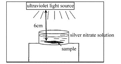
Figure 2. Schematic diagram of silver particles films growth by photocatalytic reduction method
![]()
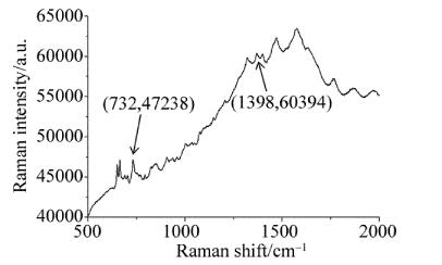
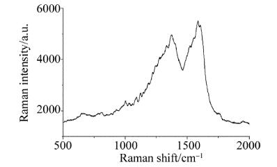
Figure 4. Surface enhanced raman spectra of aconite solution by using silver sol nanoparticle solution as substrate
-
[1] YANG D, YING Y. Applications of Raman spectroscopy in agricultural products and food analysis: a review[J]. Application Specification Reviews, 2011, 46(7): 539-560.
[2] SCHLÜCKER S. Surface-enhanced Raman spectroscopy: concepts and chemical applications[J]. Angewandte Chemie—International Edition, 2014, 53(19): 4756-4795. DOI: 10.1002/anie.201205748
[3] LU Sh Y, WANG Sh G, LIU W J, et al. Raman spectroscopy in ovarian cancer diagnostics[J]. Spectroscopy and Spectral Analysis, 2017, 37(6):1784-1788(in Chinese). http://www.wanfangdata.com.cn/details/detail.do?_type=perio&id=gpxygpfx201706025
[4] LI W, FAN X G, WANG X, et al.Design of rapid detection system for urotropine in food based on SERS[J]. Spectroscopy and Spectral Analysis, 2017, 37(6):1778-1783(in Chinese). http://www.wanfangdata.com.cn/details/detail.do?_type=perio&id=gpxygpfx201706024
[5] DONG J L, HONG M J, ZHENG X Q, et al. Discrimination of human, dog and rabbit blood using Raman spectroscopy[J]. Spectroscopy and Spectral Analysis, 2018, 38(2):459-466(in Chinese).
[6] FAN Y X, LAI K Q, RASCO BARBARA A, et al. Analyses of phosmet residues in apples with surface-enhanced Raman spectroscopy[J]. Food Control, 2014, 37(1):153-157. http://www.wanfangdata.com.cn/details/detail.do?_type=perio&id=b5945c499def0126e3d2f09959a286e4
[7] OU Y M, PEI L, L K Q, et al. Rapid analysis of multiple sudan dyes in chili flakes using surface-enhanced Raman spectroscopy coupled with Au-Ag core-shell nanospheres[J]. Food Analytical Methods, 2017, 10(3): 565-574. DOI: 10.1007/s12161-016-0618-z
[8] LIU Y D, X Q H, WANG H Y, et al. Quantitative study on phosmot residues in navel oranges based on surface enhanced Raman spectra[J]. Laser Technology, 2017, 41(4): 545-548(in Chinese).
[9] SHARMA Y, DHAWAN A. Plasmonic "nano-fingers on nanowires"as SERS substrates[J]. Optics Letters, 2016, 41(9): 2085-2088. DOI: 10.1364/OL.41.002085
[10] HUANG Y, CHEN Y, XUE X T, et al. Unexpected large nanoparticle size of single dimer hotspot systems for broadband SERS enhancement[J]. Optics Letters, 2018, 43(10): 2332-2335. DOI: 10.1364/OL.43.002332
[11] LI R P, LI Y M, HAN J H, et al. In situ SERS monitoring of plasmonic nano-dopants during photopolymerization[J]. Optics Letters, 2017, 42(9): 1712-1715. DOI: 10.1364/OL.42.001712
[12] TIAN Y, ZHANG H, XU L L, et al. Self-assembled monolayers of bimetallic Au/Ag nanospheres with superior surface-enhanced Raman scattering activity for ultra-sensitive triphenylmethane dyes detection[J]. Optics Letters, 2018, 43(4): 635-638. DOI: 10.1364/OL.43.000635
[13] LIN R B, HU L, WANG J Zh, et al. Raman scattering enhancement of a single ZnO nanorod decorated with Ag nanoparticles: synergies of defects and plasmons[J]. Optics Letters, 2018, 43(10): 2244-2247. DOI: 10.1364/OL.43.002244
[14] YE Y, LIU Y, SUN S. Theoretical and experimental study on Raman spectra of ammonium thiocyanate solution[J]. Laser Technology, 2015, 39(2): 280-283(in Chinese). http://www.wanfangdata.com.cn/details/detail.do?_type=perio&id=jgjs201502028
[15] ZHENG L M, LV Y W, TANG Sh X, et al. Phase growth mechanism of ultra-fine nano-diamond prepared by nanosecond laser[J]. Laser Technology, 2016, 40(1): 25-28(in Chinese). http://www.wanfangdata.com.cn/details/detail.do?_type=perio&id=jgjs201601007
[16] DENG Y.Comparative study of three SERS active substractes based on AgNPs[D]. Dalian: Dalian University of Technology, 2015: 16-36(in Chinese).
[17] LEE P C, MEISEL D. Adsorption and surface-enhanced Raman of Dyes on silver and gold sols[J]. The Journal of Physical Chemistry, 1982, 86(17): 3391-3395. DOI: 10.1021/j100214a025
[18] JI Sh F, JIANG T L, XU K, et al. FTIR study of the adsorption of water on ultradispersed diamond powder surface[J]. Applied Surface Science, 1998, 133(4): 231-238. http://www.wanfangdata.com.cn/details/detail.do?_type=perio&id=44f881799f336b2d3bbe00d514d2e684
[19] LIU Y, LIU Ch Y, ZHANG Zh Y, et al. The surface enhanced Raman scattering effects of composite nanocrystals of Ag-TiO2[J]. Spectrochimica Acta, 2001, A57(1):35-39. http://www.wanfangdata.com.cn/details/detail.do?_type=perio&id=c872098f56ea37ff71ebeb42e6c4d7eb
[20] YANG H D, LIN X, LIU Y L, et al. Preparation of three-dimensional hotpot SERS Substrate with silver nanocubes and its application in detection of pesticide[J]. Spectroscopy and Spectral Analysis, 2018, 38(1): 99-103(in Chinese). http://www.wanfangdata.com.cn/details/detail.do?_type=perio&id=gpxygpfx201801020
[21] LI D W. Controlled synthesis of carbon nanocoils and their application in SERS[D]. Dalian: Dalian University of Technology, 2013: 87-98(in Chinese).
 下载:
下载:
计量
- 文章访问数: 5
- HTML全文浏览量: 0
- PDF下载量: 2